The Optic Disc as the Basis for Effective Treatment of Glaucoma
Dr.Rezaul Murshed
M.B.B.S(D.M.C).D.O(D.U)
AHMAD MEDICAL CENTER LTD
71, Dhanmond Residential Area
Road.#15A, Dhaka-1209
Cell: 0171-533357 Res: +880-2-8152761
Optic Disc
Optic Nerve Examination
Optic Nerve Characteristics
Optic Nerve Size and Glaucoma
The Normal Nerve - Neuroretinal Rim Shape
--The ISNT rule (Jost Jonas)
--Thickest rim Inferiorly > Superiorly > Nasally > Temporally (Thinnest)
--Critical to examine in early glaucoma
SIMPLE DRAWING

I:S = 2:1.5, N:T= 2:1
Neuroretinal Rim Shape and Glaucoma
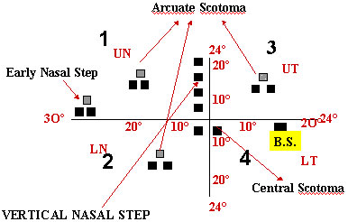
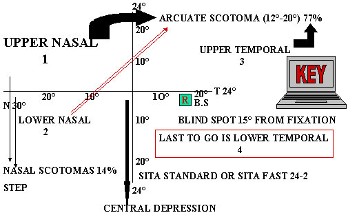
--First loss is typically inferotemporal and then superotemporal
--For more advanced disease temporal regions are more thinned
--For very advanced disease typically the nasal rim is the thickest remaining part (superonasal thicker)
--Typically affect upper nasal quadrant first
--Then inferior field and so on
--End stage disease often has only a inferior temporal island left (superonasal rim)
--Larger and more laminar pores at the poles might be important
--Laminar bowing is more prominent at the poles
--Axons are more kinked at the disc periphery
Advance glaucoma


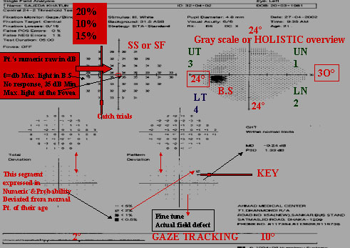
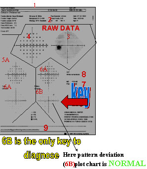
This school teacher of 45yrs. enjoys 6/4.5R/E&; 6/6 L/E. Binoculr Esterman shows 4 adjacent scotoms in the
Sup. Field & 6 scotomas seen in the lower Temp. zone. Here out of 120 points, he can see 109 & cannot See 11 points. When not seen crosses 10>in screening test than full Threshold test to be done. This is an advance case of POAG R>L.
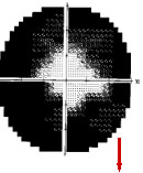
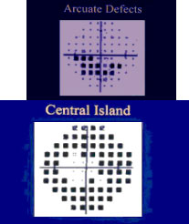
DENSE SUP ARCUATE SCOTOMA

DENSE SUP ARCUATE SCOTOMAS EMANTING FROM THE BLIND SPOT JOINS THE SUP ARCUATE SCOTOMA LATER JIONS THE NASAL SUP NASAL STEP.FIELD MATCHES WITH THIN INF NRR.A CASE OF ESTABLISHED POAG.
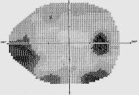
DISC SHOWS NO INFERIOR NEURO RETINAL RIM
NORMAL NERVE-NRR SHAPE
Asymmetric DISC
Tissue between The cup & disc is NRR made of nerve & capillaries So, looks red to orange
NRR
Cup size and Disc Size
Optic Nerve Head Cupping is a Unifying Feature of all GLAUCOMAS
Cupping

Enlargement of cup / loss of Neuroretinal Rim
Disc Examination
OLD IS GOLD

Optic Nerve Examination Problems
HORIZONTAL C:D 0.4, CUP OCCUPIES 4/10 WIDTH OF DISC "Armaly"

Problems with Armaly's c/d
Healthy Disc
c/d 0.5
Normal Field
Sick disc

c/d 0.5
Field defect
Disc Size
Small disc
1.0 mm
c/d 0.2Disc Size <1.3mm
Large disc

2.Omm
c/d 0.6 Disc size 2mm
Here Smaller disc is more damaged
Determine Thinnest Rim/Disc
Ratio
Rim Disc/Disc 0.2
Here thinnest rim .2mm & if disc size 1 than Rim/Disc = 0.2
Normal field

Rim less than <45
Field defect
Optic Nerve Examination Problems
Which of these nerves is more likely Pathologic?

The Disc Damage likelihood Damage Scale (DDLS)
Spaeth G & Henderer J 2002 May
The Disc Damage Likelihood Scale (DDLS)
Comparison of cup/disc and Rim/Disc ratio

Cup/Disc = 0.1
Rim/Disc = 0.45

C/Disc = 0.3
Rim/Disc = 0.35

Cup/Disc = 0.8
Rim/Disc = 0.1

Cup/Disc = 0.9
Rim/Disc = 0.05
When disc is arbitrarily Considered 1
BY SLIT LAMP

Conversion Chart
| Manufacturer | LENS | ||
| 60D | 78D | 90D | |
| VOLK | 0.88 | 1.11 | 1.33 |
| NIKON | 1.03 | 1.63 |
Lim, et al. J Glaucoma 1996; 5:241
Measuring Disc Diameter
-L ens specific
- Manufacturer specific
Example
1.6mm(Vertical length)x1.11(Volk 78)=1.78mm, will be the disc diameter.
How to determine the rim/disc ratio

So, rim/disc=.35/1.78=0.19
Stage 2
When damage will occur
Theoretical R im Area by Optic Nerve Diameter
Henderer 2000
Estimation of Disc Pathology by Rim/Disc ratio
DDLS STAGING

a is a healthy optic disc, DDLS Stage Oa, rim/disc .4
b is a possible disc damage DDLS Stage 2, rim/disc .25
c is a severely damaged optic disc,stage 7a, no rim From 9.30 to 5.30
b is a possible disc damage DDLS Stage 2, rim/disc .25
c is a severely damaged optic disc,stage 7a, no rim From 9.30 to 5.30
Staging of DDLS
The Disc Damage Likelihood Damage Scale
| STAGE |
Narrowest Width of rim or Circumferential no rim |
| 0 | 0.3-0.5 |
| 1 | 0.2-0.29 |
| 2 | 0.1-0.19 |
| 3 | 0.01-0.1 |
| 4 | No rim <45 ° |
| 5 | No rim 45°-90° |
| 6 | No rim 91°-180° |
| 7 | No rim > 180° |


DDLS Conclusions
DDLS not the Key
PROVIEW TONOMETER
